
If your spine surgeon suspects that a specific condition may be causing your pain, they will order diagnostic tests. These could include imaging, scans or blood tests. At Huffman Clinic’s spine surgery center, we perform a complete set of tests to ensure that our doctors understand every aspect of your health and pain before choosing a treatment.
X-ray
An X-ray is a machine that takes a picture of your bones. While the photos are being taken, you’ll be asked to lie still in different positions so the machine can capture specific areas. The process is painless, but if you feel any discomfort you should tell the technician. In the case of spinal pain, your doctor may order an X-ray to observe a few different aspects of your spine including:
- The distance between discs
- Bone spurs
- Nerve bundle sclerosis (hardening)
- Facet hypertrophy (enlargement)
- Instability during flexion or extension of limbs.
Because an X-ray can only show bones, your spine surgeon will most likely use it in conjunction with another test.
MRI Scan (Magnetic Resonance Imaging)
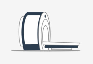
Similar to an X-ray, the MRI is used to take images of the inside of your body. However, the MRI is able to cut through multiple layers of the spine and show any abnormality of soft tissues, such as nerves and ligaments. This is done using a magnetic field. During the scan, you will lie on a long table that slides into the MRI machine shaped like a tunnel. The process is painless make sure to tell your technician if you feel discomfort or claustrophobic. The total procedure typically takes about 30-60 minutes. During an MRI, your doctor may look for:
- Loss of water in a disc
- Facet joint hypertrophy (enlargement)
- Stenosis (narrowing of the spinal canal)
- A herniated disc (protrusion of the intervertebral disc).
CT/CAT Scans (Computer Assisted Tomography)
The CAT scan is used to show both bones and soft tissues. To do this, the machine takes a set of cross-sectional images to show disc problems and degeneration of bones. Imagine a loaf of bread. The images a CT/CAT scan takes are like slices. When the slices are put together, they form a loaf. When the CT/CAT scan images are put together, they form a 3D picture of your spine.
Similar to an MRI, during a CAT scan, you will lie on a table that slides into a scanner. The scanner rotates in a circle taking many pictures, which may sound like loud clicking noises. The procedure takes 30-60 minutes.
Myelogram
A myelogram uses a special dye to outline the spinal cord and nerve roots during an X-ray. Before a myelogram, you’ll need to be injected with the special dye. Your doctor will insert a small needle into the spinal canal. This process is called a spinal tap.
Once you’ve had the spinal tap, you’ll lie on a table, which will begin to tilt in order to move the dye around your spinal sac. The movement of the dye is captured by an X-ray machine. Your spine surgeon will observe this movement to understand abnormalities in the shape of your spinal sac.
Bone Scan
A bone scan uses a radioactive chemical, sometimes called a tracer, to identify hotspots in the bone structure. A hotspot is an area where there’s a lot of radioactivity. Your doctor can use these hotspots to understand where your pain is coming from and where to look next.
The tracer is injected into the bloodstream through an IV, where it will attach itself to any hotspot areas. A special camera will take pictures of your skeleton over the period of time the tracer is in your body. Hotspots will show up as dark areas on the images because the chemical tracer will accumulate in the area and the radiation will be picked up by the camera.
Your doctor may order a bone density test if they’re concerned about osteoporosis.
EMG/SSP (Electro-diagnostic Study)
An electromyogram (EMG) looks at the function of your nerve roots leaving the spine, by inserting tiny electrodes into your muscles. The electrodes can identify abnormal signals in the muscles which indicates if a nerve is being irritated or pinched as it leaves the spine. If the EMG machine finds that muscles are not working properly, your doctor can assume that the nerves must be getting pinched somewhere.
Facet Joint Block
The facet joints are the joints in your spine that make it easy to move. If they’re irritated or inflamed, they can cause back pain. The facet joint block is a procedure where a local anesthetic medication is injected into the facet joint to numb the area around it. If all your pain goes away, your doctor will know that your spinal pain was caused by issues in your facet joints.
Discogram
A discogram is an X-ray procedure that looks at the discs in your spine. The test is conducted by injecting dye into the center of the injured disc to make it clearly visible on film and screen. Before the procedure begins you will be given local anesthetic and put to sleep. A discogram usually lasts about 40 minutes.
Labs
Your doctor may order a complete blood count, inflammatory markers, urine analysis, fecal occult blood testing or other labs.
Further lab tests may be done to check for problems not related to the deterioration of the spine. Testing can help determine the presence of serious problems, such as infection, arthritis, cancer, or an aortic aneurysm.
Spinal Tap
During a spinal tap, a needle is inserted into your spinal canal. Spinal taps are conducted to get a sample of the clear fluid that surrounds your spinal cord. While its composition is similar to blood, the fluid typically does not contain red blood cells or many white blood cells. With this, the presence of an increased count of white blood cells, an increase in protein level, or inflammation can indicate an issue in the spine.